

Isolation and Identification of Duck Plague Virus
-
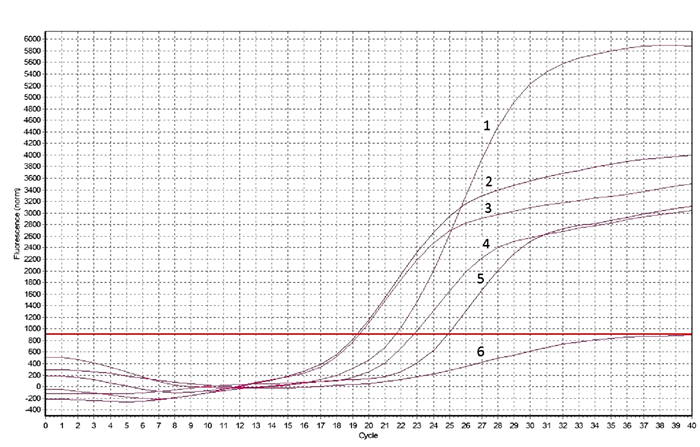
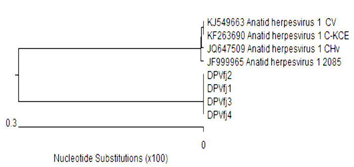
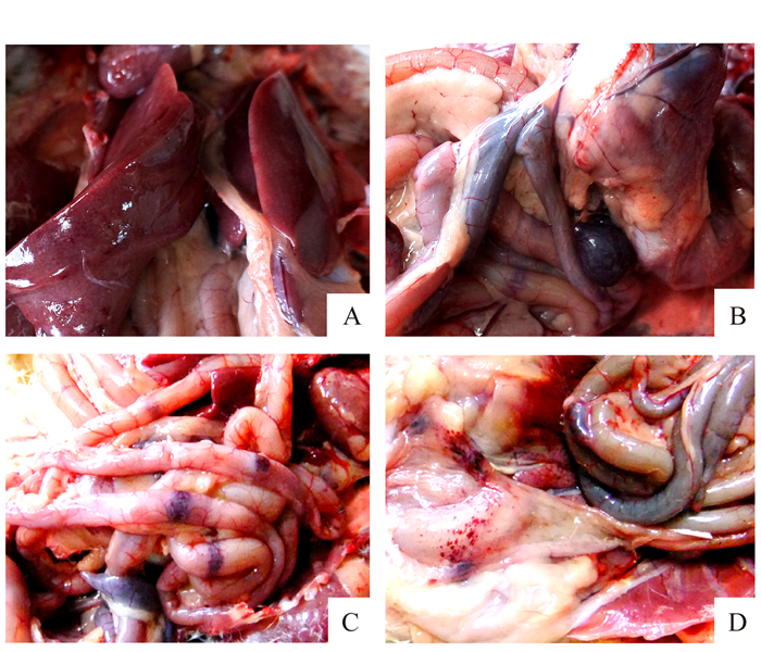
摘要: 采集疑似鸭瘟病毒自然感染的病死番鸭的肝脾等组织,应用番鸭胚成纤维细胞(MDEF)进行病毒分离,通过对分离毒的血凝特性(HA)测定、间接免疫荧光试验(IFA)荧光定量PCR、PCR产物测序和动物回归试验等初步鉴定。结果显示:通过MDEF从疑似病料中分离到4株病毒(DPVfj1、DPVfj2、DPVfj3、DPVfj4),均不能凝集鸽红细胞;IFA结果排除了分离毒为鹅细小病毒、番鸭细小病毒、鸭呼肠孤病毒、鸭副粘病毒和禽坦布苏病毒;鸭瘟病毒荧光PCR试剂盒检测分离毒核酸均为鸭瘟阳性;鸭瘟病毒(JQ673560)gJ蛋白基因序列特异引物进行PCR扩增均为阳性,且PCR产物序列与鸭瘟病毒参考株gJ蛋白基因序列相似度均大于99%;动物回归试验显示,分离毒人工感染30日龄番鸭和同居感染5日龄雏番鸭均可复制出与自然感染一致的临床表现及病理变化,并能回收到病毒。上述结果表明4株分离毒均为鸭瘟病毒强毒株。Abstract: Tissue samples of liver and spleen from the Muscovy ducks that were suspected to have died from the duck plague were collected for this study.Viruses were isolated from the Muscovy duck embryo fibroblasts (MDEF) for a preliminary identification using hemagglutination assay(HA), immunofluorescence analysis(IFA), qPCR, PCR product sequencing, and animal infection test. Four virus strains, DPVfj1, DPVfj2, DPVfj3 and DPVfj4, were thus identified for further investigation. Subsequently, it was found that these strains (a) would not cause red blood cell agglutination on pigeon erythrocytes; (b) were tested negative on IFA with goose parvovirus, Muscovy duck parvovirus, duck reovirus, duck paramyxovirus, and Tembusu virus; (c) showed positive on test with fluorescence RT-PCR kit; (d) had a positive result on their PCR-amplified pair of primers from the specific fragment of gJ protein gene, JQ673560, and a homologygreater than 99% on the sequence of the fragment with that of the duck plague virus(DPV) gJ protein gene; (e)infected 30-day-old ducks by injection and 5-day-old ducklings by co-habitation showing exactly the same clinical symptoms and postmortem signsas in the natural cases; and, (f)were recovered from the diseased birds. The results seemed to verify the fact that DPV caused the duck plaque in natural environment, and that the isolated strains possessed the virulent nature in question.
-
Keywords:
- duck plague virus /
- isolation /
- identification
-
-
![]()
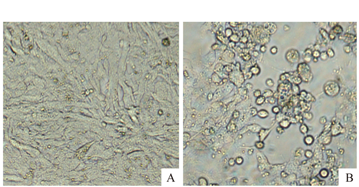
图 1 分离毒致MDEF单层细胞的CPE
注:A为MDEF对照;B为接毒后72 h CPE。
Figure 1. CPE of MDEF monolayer on cells infected by isolated viruses
-
[1] DAVISON S, CONVERSE K A, HAMIR A N, et al.Duck viral enteritis in Muscovy ducks in Pennsylvania[J].Avian Dis, 1993, 37:1142-1146. DOI: 10.2307/1591927
[2] 殷震, 刘景华.动物病毒学:第2版[M].北京:科技出版社, 1997:1073-1077. [3] ARAVIND S, NITIN M K, SATISH S G, et al. Adaptation and growth kinetics study of an Indian isolate of virulent duck enteritis virus in vero cells[J]. Microbial Pathogennesis, 2015, 78:14-19. DOI: 10.1016/j.micpath.2014.11.008
[4] 甘孟侯.中国禽病学[M].北京:中国农业出版社, 1999:107-119. [5] 魏笑笑, 高亚东, 刘志杰, 等.一株鸭瘟病毒GM株的分离与鉴定[J].中国家禽, 2015, 37(6):63-65. http://www.cnki.com.cn/Article/CJFDTOTAL-ZGJQ201506020.htm [6] 王红海, 胡薛英, 苏敬良, 等.商品肉鸭鸭瘟病毒的分离与鉴定[J].中国预防兽医学报, 2006, 28(1):105-108. http://www.cnki.com.cn/Article/CJFDTOTAL-ZGXQ200601028.htm [7] 马秀丽, 宋敏训, 艾武.山东省鸭瘟病jh株的分离鉴定[J].中国畜牧兽医, 2005, 32(9):47-49. http://kns.cnki.net/KCMS/detail/detail.aspx?filename=gwxk200509017&dbname=CJFD&dbcode=CJFQ [8] 孟刚, 王经满, 曹瑞兵.2株鸭瘟病毒强毒株的分离鉴定[J].畜牧与兽医, 2012, 44(4):59-62. http://www.cnki.com.cn/Article/CJFDTOTAL-XMYS201204019.htm [9] 付书林, 陈夏冰, 钱运国, 等.一株鸭瘟病毒的分离与鉴定[J].湖北农业科学, 2014, 53(19):4644-4646. http://www.cnki.com.cn/Article/CJFDTOTAL-ZGJQ201506020.htm [10] 马圣利.一株鸭瘟病毒的分离与鉴定[J].现代农业科技, 2016, (8):261-262. http://www.cnki.com.cn/Article/CJFDTOTAL-ZGJQ201506020.htm
 下载:
下载:
计量
- 文章访问数: 1349
- HTML全文浏览量: 289
- PDF下载量: 248
